- Ophthalmologist – Unlike other eye care team members, ophthalmologists are medical doctors (MDs). They usually are responsible for advanced eye care and eye surgeries.
- An optometrist isn’t a medical doctor and is responsible for routine eye and vision care.
- Optician – A person who fits contact lenses and eyeglasses by fulfilling a prescription issued by an ophthalmologist or optometrist.
- Slit lamp exam
- Visual acuity test
- Intraocular pressure test
- Visual field
- Fundoscopy
- Confrontation visual field test: Your doctor asks you to look at the point directly before you, such as the doctor’s nose. One of your eyes is covered, and the specialist shows you a different number of fingers in the side vision fields.
- Automated static perimetry test – A machine creates a specific map of where you can and can’t see. To do so, you look into the center point of the machine, and a small dim of light appears in various places. When you notice the light, you press a button. The machine then creates a map of which spots you saw and which you didn’t.
- Glaucoma
- Cataracts
- Age-related macular degeneration (AMD)
- Dry eye
- Pink eye
- Retinal detachment
- Lazy eye
- Stye
- Color blindness
- Floaters
- Dry – occurs in 80-90% of patients; it has a milder course;
- Wet – develops more dynamically and is usually associated with more serious visual impairment.
- To stop the development of dry AMD, follow an appropriate diet. You can also use supplements.
- In the treatment of the wet form, you will have to undergo injections in the eyeball performed under local anesthesia.
- American Academy of Ophthalmology (AAO) What Is an Ophthalmologist vs Optometrist? (2024) https://www.aao.org/eye-health/tips-prevention/what-is-ophthalmologist
- National Library of Medicine (NIH) Harrison F. Daiber; David M. Gnugnoli. Visual Acuity (2023) https://www.ncbi.nlm.nih.gov/books/NBK563298/
- American Academy of Ophthalmology (AAO) Visual Field Test (2022)
https://www.aao.org/eye-health/tips-prevention/visual-field-testing - National Eye Institute (NIH) At a glance: Cataracts (2023)
https://www.nei.nih.gov/learn-about-eye-health/eye-conditions-and-diseases/cataracts - National Eye Institute (NIH) At a glance: Glaucoma (2023)
https://www.nei.nih.gov/learn-about-eye-health/eye-conditions-and-diseases/glaucoma - American Academy of Ophthalmology (AAO) What Is Macular Degeneration? (2023) https://www.aao.org/eye-health/diseases/amd-macular-degeneration
- National Eye Institute (NIH) At a glance: Dry Eye (2023)
https://www.nei.nih.gov/learn-about-eye-health/eye-conditions-and-diseases/dry-eye - National Eye Institute (NIH) At a glance: Retinal Detachment (2023)
https://www.nei.nih.gov/learn-about-eye-health/eye-conditions-and-diseases/retinal-detachment - American Academy of Ophthalmology (AAO) Conjunctivitis: What Is Pink Eye? (2023) https://www.aao.org/eye-health/diseases/pink-eye-conjunctivitis
- Kaur S, Sharda S, Aggarwal H, Dadeya S. Comprehensive review of amblyopia: Types and management. Indian J Ophthalmol. 2023 Jul;71(7):2677-2686. doi: 10.4103/IJO.IJO_338_23. PMID: 37417105; PMCID: PMC10491072.
https://www.ncbi.nlm.nih.gov/pmc/articles/PMC10491072/ - Wolvaardt E, Stevens S. Measuring intraocular pressure. Community Eye Health. 2019;32(107):56-57. Epub 2019 Dec 17. PMID: 32123477; PMCID: PMC7041827. https://www.ncbi.nlm.nih.gov/pmc/articles/PMC7041827/
- National Eye Institute (NIH) At a glance: Amblyopia (2022)
https://www.nei.nih.gov/learn-about-eye-health/eye-conditions-and-diseases/amblyopia-lazy-eye
To become an ophthalmologist, an individual has to complete many more years of training than the rest of an eye team. Therefore, an ophthalmologist is the most qualified among eye professionals and can manage a broader range of vision and eye problems.
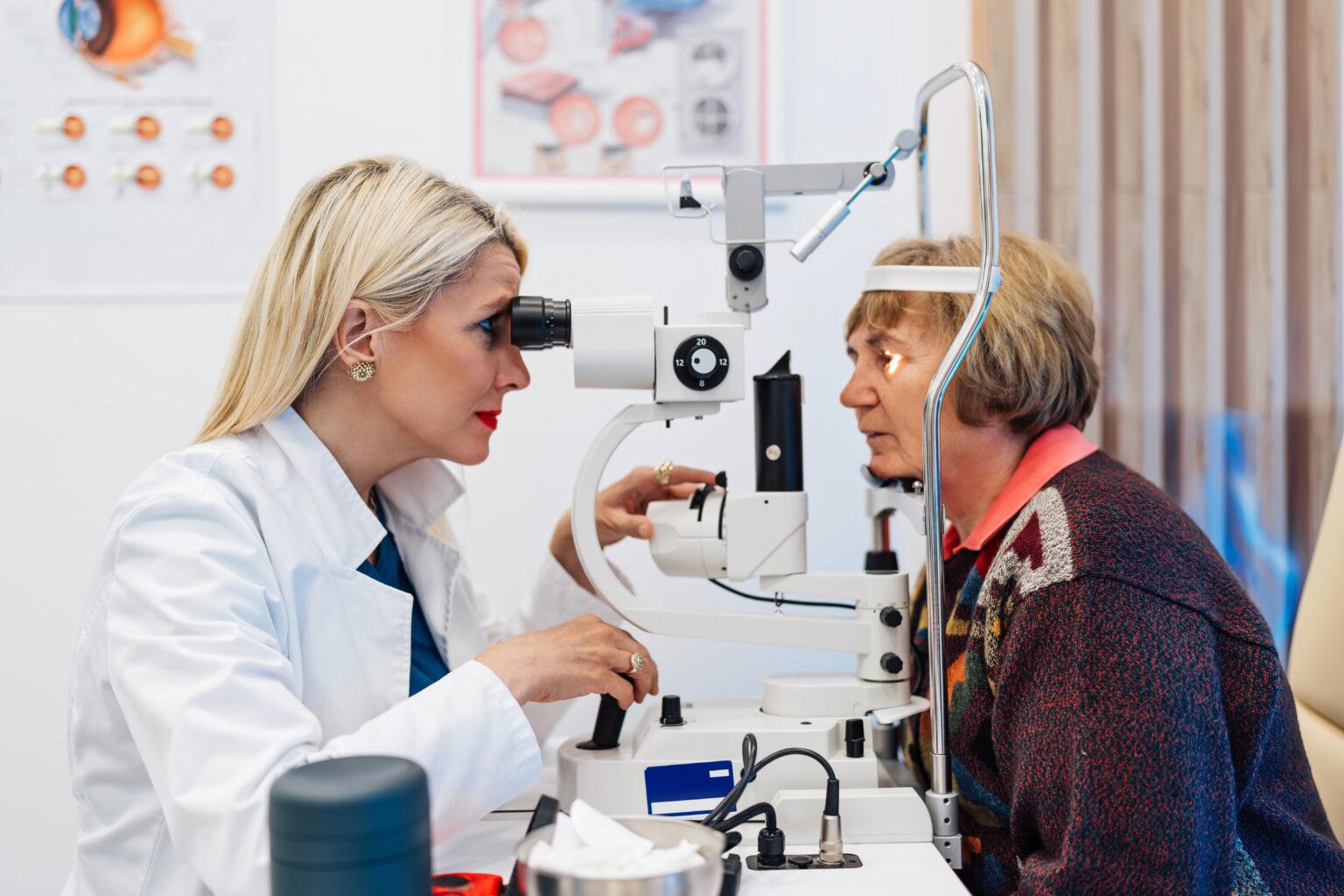
Eye Exam
An ophthalmologist performs an eye exam, which consists of several parts. Those include:
Not all the parts are performed each time you visit an eye doctor since the specialist matches the proper test to your problem. Every test provides crucial information about your eye’s specific part or function.
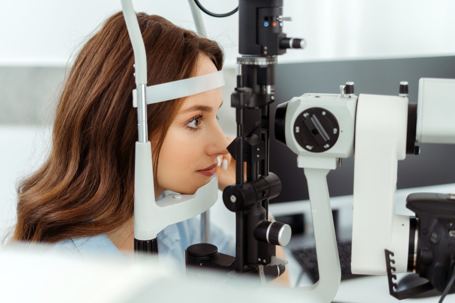
Slit Lamp Exam
The slit lamp is a special microscope with a bright light source that enables an ophthalmologist to visually assess all parts of the eye, including the inside of them.
During the examination, the doctor often dilates your pupil to see inside your eye, which causes blurred vision and light sensitivity, which disappear after a few hours, so it is worth bringing sunglasses to the examination. The head and chin should be rested on the slit lamp support throughout the examination. It would be best if you did not change the position of the head because even a slight movement causes the image in the slit lamp to become blurry.
Visual Acuity
The visual acuity test measures how well you see. It is a quick way to determine how well your vision is and whether it has changed. The most common type of this test is performed using a Snellen chart, a whiteboard with printed letters that gradually get smaller. Your specialist asks you to read the letters from a distance until you can’t tell them apart. For children, unique Snellen charts are made, which, instead of letters, display various shapes.
After performing such a test, your eye specialist may prescribe eyeglasses or contact lenses if the test shows that you need them.
Visual acuity may also be checked for near vision. To do that, your provider may ask you to hold the near vision board about 14 inches from your face and read the letters.
Intraocular Pressure Test
Inside your eyes is a fluid with a specific pressure called intraocular (eye) pressure. An eye specialist measures it in millimeters of mercury (mmHg). Eye pressure is an essential measurement since too high intraocular pressure leads to glaucoma development, which may even lead to blindness.
The test for intraocular pressure is called tonometry. The most common type is contactless air-puff tonometry, in which a machine blows a puff of air toward the eye and measures its pressure. A correct eye pressure should be between 10 mmHg and 21 mmHg.
Visual Field
Visual field means how wide you can see when focusing on the point in front of you. There are several types of visual field tests (perimetry). The most commonly used are:
Fundoscopy
In fundoscopy, an ophthalmologist uses a unique device to examine the back of the eyeball and assess the retina, blood vessels, and optic nerve. The retina is a layer of the eye that converts light into neural signals, creating an image.
Conditions
Ophthalmologists diagnose and treat various conditions affecting the eyes and vision. The most common ones include:
Glaucoma
Glaucoma is a group of diseases most often characterized by increased intraocular pressure and, if left untreated, the gradual damage of the optic nerve, i.e., the path connecting the retina with the brain. Optic nerve damage results primarily in defects in the field of vision and may lead to complete loss of vision.
Depending on the stage of the disease, the vision range of glaucoma patients may gradually shrink, leading to so-called tunnel vision. This means that central vision remains intact, but peripheral vision (the perception of objects not directly in front of the eyes) is significantly reduced. In glaucoma, there are also difficulties in distinguishing colors and contrasts.
Glaucoma treatment involves controlling the pressure inside the eyeball and stopping the progression of the disease. Several treatments can be used alone or in combination. Ophthalmologists often recommend medications (in the form of eye drops) that lower the pressure inside the eyeball. When normalizing eye pressure becomes difficult, your ophthalmologist may consider laser therapy to improve fluid outflow from the eyeball. The same effect can also be achieved through surgery.
Cataracts
Cataract is a disease that causes the eye’s lens to become cloudy. A healthy lens is perfectly transparent, which leads to good vision quality. As a result of this disease, its surface loses its transparency, and the patient suffering from cataracts has worse vision. As a result of clouding, the lens allows much fewer rays to pass through than a healthy lens, which is why it results in a blurred image, much weaker color saturation, and reduced contrast. This contributes to a significant reduction in visual comfort.
There are several types of the condition, the most common being age-related. In the early stages of this type of disease, an ophthalmologist may prescribe anti-cataract eyedrops.
If the cataract has affected a large eye surface or is already in a more advanced stage, surgery is often necessary. It involves removing the damaged lens and replacing it with an artificial one.
Congenital cataracts are treated only surgically. This procedure should be performed as early as possible so the child can develop properly.
Age-Related Macular Degeneration
In age-related macular degeneration (AMD), a part of the retina, the macula, is destroyed. As a result, central vision deteriorates while side vision remains normal. AMD is a common condition and a leading cause of vision loss in people over 50. There are two kinds of AMD:
Adequately conducted therapy will help you reduce symptoms and slow down the progression of the disease.
The ophthalmologist will select the treatment depending on the form of the disease:
Dry Eye
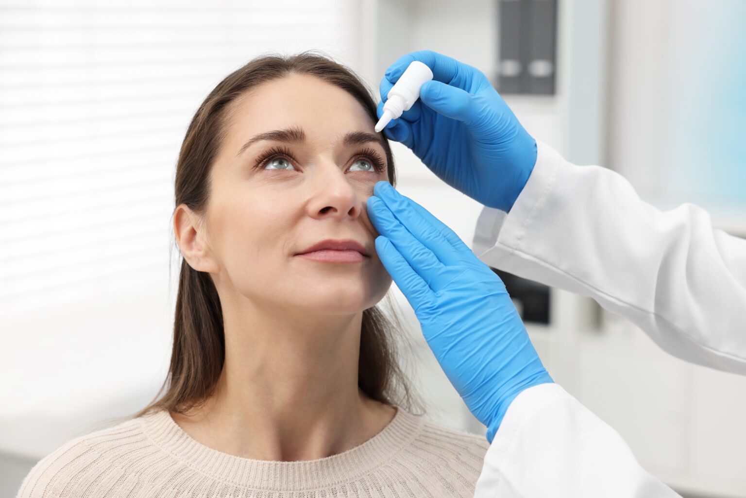
Dry eye develops when your eyes don’t produce enough tears or work correctly. Too few tears cause the eyes not to stay wet, leading to discomfort, a scratchy feeling in the eyes, blurry vision, and red eyes.
Thankfully, there are many treatment options for uncomfortable symptoms. These include lifestyle changes, over-the-counter moisturizing eye drops, prescription medications, tear duct plugs, and surgery.
Pink Eye
Pink eye is another name for inflammation of the conjunctiva (conjunctivitis), the transparent layer of tissue in front of the eye. During this condition, your eye becomes red and swollen, and a sticky discharge develops. The condition can be contagious (when bacteria, viruses, or fungi cause it) or not contagious (when caused by allergies, injuries, and other factors). Depending on the cause, an ophthalmologist treats pink eye with antibiotics, allergies, and antiviral medications. Some types of pink eye pass on their own, and then alleviating symptoms with a cold washcloth, painkillers, and anti-inflammatory medications are recommended.
Retinal Detachment
Retinal detachment occurs when the retina (the thin layer of tissue at the back of the eye) detaches from the surrounding tissues. It is a painless but severe condition that can affect vision and may lead to blindness. The detached retina stops getting nutrients and oxygen from the blood vessels, which is crucial to correct functioning.
The symptoms of retinal detachment include seeing flashes of light, having more floaters than usual (dark spots that drift across the vision), and darkening of your side vision. An ophthalmologist can diagnose retinal detachment during an eye exam. Treatment options include laser therapy and surgery.
Lazy Eye
A lazy eye – or what some doctors call amblyopia – is when someone struggles to see clearly out of both eyes simultaneously. It usually happens to infants and little children, frequently related to their eyes not lining up quite right. Think about it like this: they can look through one eye perfectly well, but everything turns blurry when they try simultaneously using the other. When both eyes don’t provide the same vision, the brain ignores the vision from one of the eyes, leading to the lazy eye. If left untreated, the condition may lead to vision loss in one eye.
An ophthalmologist treats amblyopia. Treatment options include wearing an eye patch, eyeglasses, and eye surgery.
When Should You Visit Ophthalmologist?
If you notice any troubling symptoms related to your eye health, you should visit your ophthalmologist immediately. Those symptoms, including blurry vision, worsening of vision during nighttime, double vision, cloudy vision, eye redness, and abnormal eye discharge, should be addressed immediately.

