During reproduction, an individual inherits one chromosome from each parent, with chromosomes serving as carriers of genes, the fundamental units of heredity. Consequently, chromosomes play a pivotal role in the genetic transmission and determine various facets of an organism’s development, functionality, and behavioral, and physical traits.
Structure
The structure of a chromosome is simple, although it varies depending on the stage of cell division. The chromosome consists of two arms, which are maximally shortened in prophase. In subsequent phases, it elongates until it finally forms the shape known from descriptions and drawings – its arms form twin chromatids that connect at the centromere. In this form, the chromosome is at the metaphase stage, i.e., just before cell division.
What it contains is also significant. The human chromosome consists of:
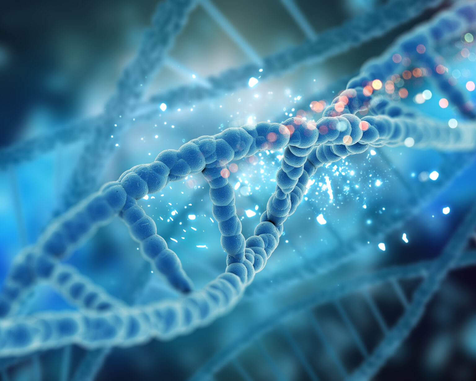
- Deoxyribonucleic acid, or DNA – is a thin and long molecule that constitutes a set of genes and, therefore, information on how to code individual amino acids; DNA from one chromosome would be several centimeters long when stretched
- Histones, i.e., proteins that constitute the structure around which DNA is wound and condensed
- Nucleolar organizer, i.e., secondary constriction (in acrocentric chromosomes)
Chromatin is the structure in which double-stranded DNA is wound around histone protein molecules. Thanks to it, the chromosome is a safe hiding place for genetic material.
Types
There are several types of chromosomes based on their structure.
Acrocentric chromosomes are characterized by a short arm with a secondary constriction containing nucleolar organizer sequences for encoding nucleoli. The centromere is near the end of the brief ‘p’ arm, and examples of acrocentric chromosomes include chromosomes 13, 14, 15, 21, and 22.
Metacentric chromosomes have a centromere positioned at the chromosome’s center, resulting in arms of equal length forming a V shape. Submetacentric chromosomes have a centromere close to, but not at, the center, resulting in an L shape during metaphase and anaphase. Telocentric chromosomes have only one arm with the centromere at the end and are absent in humans.
Homologous chromosomes in humans consist of almost identical pairs, possessing the same shape and size and similar genetic information. When these chromosomes differ in form, they are called alleles, leading to a heterozygous state. Conversely, the cell is termed homozygous when these chromosomes share the same gene form. Homologous chromosomes make up autosomes, which are non-sex chromosomes.
Homologous chromosomes comprise a pair of chromosomes, one from the mother and the other from the father. Pairing occurs briefly during meiosis. Then, homologous chromosomes spread to opposite poles of the dividing cell. It is how a gamete is created, consisting of cells with a haploid chromosome number.
Sex Chromosomes
There is a 23rd pair of allosomes in the chromosome. The allosome is the chromosome that determines sex. Both men and women have one X chromosome, which is the same, but the other one varies.
The X chromosome is always inherited from the mother. In females, the second X chromosome from the father determines their sex as female, resulting in a system of XX chromosomes. Conversely, the Y chromosome determines the male sex. In males, one chromosome (X) comes from the mother and the other (Y) from the father, resulting in XY chromosomes.
Inheritance of the Y or X chromosome from the father does not occur when the reproductive cells (gametes) of the parents unite but during the process of meiosis of these cells. During this division, an X or Y chromosome is transferred to the gamete one at a time. Compared to other body cells, reproductive cells have 22 autosomes and only one chromosome – X in women and X or Y in men.
Chromosome Aberrations
Chromosomal aberrations are one of the most common causes of genetic diseases, spontaneous miscarriages, or intellectual disabilities. If a child with a chromosomal defect survives pregnancy, it is usually born with visible body deformities or functional disorders of internal organs.
Chromosomal aberrations are categorized into various types.
Structural Aberrations
Structural chromosomal aberrations occur due to breaks in the continuity of a chromosome or chromatid in one or two locations. This category includes:
- Inversions: when segments of a chromosome are reversed by 180° and reattached
- Translocations: the movement of a fragment from one chromosome to another
- Duplications: involving the doubling of a specific section of a chromosome
- Deletions: the loss of a specific section of a chromosome
- Insertions: the addition of a chromosome fragment into another place on the same or various chromosome
- Dicentric chromosomes: containing two centromeres
- Circular chromosomes: formed as a result of breaks in the end parts of the chromosome arms and subsequent joining of its ends
Numerical Aberrations
Numerical chromosomal aberrations result from the incorrect division of pairs of homologous chromosomes or sister chromatids during meiosis:
- Aneuploidies: an increase or decrease in the number of chromosomes by single units
- Polyploidy: the multiplication of the entire set of human chromosomes
Causes of Chromosomal Aberrations
Chromosome aberrations may occur due to spontaneous errors during the division of chromosomes into cells or as a result of mutagenic factors such as ionizing, ultraviolet, or low-energy electromagnetic radiation, toxic chemicals (alkylating compounds), radiotherapy, high temperature, certain bacterial infections or viruses, cytostatics, reactive oxygen species, and free radicals.
Diseases
Genetic diseases result from an incorrect structure or several genes or chromosomes. Some of them are inherited, while others result from new gene mutations or changes in chromosome structure due to improper distribution of autosomes in the parent’s gamete and, consequently, modification of their number in the child’s body. Genetic diseases are congenital and are diagnosed in the fetal phase or instantly after birth.
The most prevalent disorders resulting from genetic abnormalities involving genes or chromosomes are as follows.
Down Syndrome
Down syndrome is caused by the existence of an additional copy of chromosome 21, with an incidence of approximately 1 in 650-700 live births. Surviving individuals may expect a lifespan of 50-60 years, exhibiting infertility in males and a 50% chance of passing on the abnormal chromosome in females.

Edwards Syndrome
Edwards syndrome arises from an additional chromosome 18, often detected in pregnancies with indicators such as reduced fetal movement, placental underdevelopment, or fetal growth restriction.
Patau Syndrome
Patau syndrome is caused by an extra chromosome 13, characteristically rare due to high rates of miscarriage, and associated with a mortality rate of nearly 90% in infants under one year of age.
Prader-Willi Syndrome
Prader-Willi syndrome is a rare genetic condition originating from the partial loss of chromosome 15 inherited from the father. Recognizable at birth, the syndrome presents distinct physical features, reduced muscle tone, behavioral irregularities, and aggressive tendencies.
Angelman Syndrome
Angelman syndrome results from the loss of a specific segment of chromosome 15 inherited from the mother, slightly more prevalent and colloquially referenced as the “happy puppet” syndrome due to episodes of unprovoked laughter.
Williams Syndrome
Williams syndrome arises from the deletion of chromosome 7, affecting 1 in 20,000 births. It is characterized by distinct facial features, a mild temperament, and mild cognitive impairment, often accompanied by absolute pitch.
Fragile X Syndrome
Fragile X syndrome is a genetic disorder caused by a dynamic mutation (duplication of a gene segment) in the FMR1 gene located on the X chromosome. It is more commonly manifested in males and is characterized by varying degrees of intellectual impairment, often akin to autism.
Diagnostics
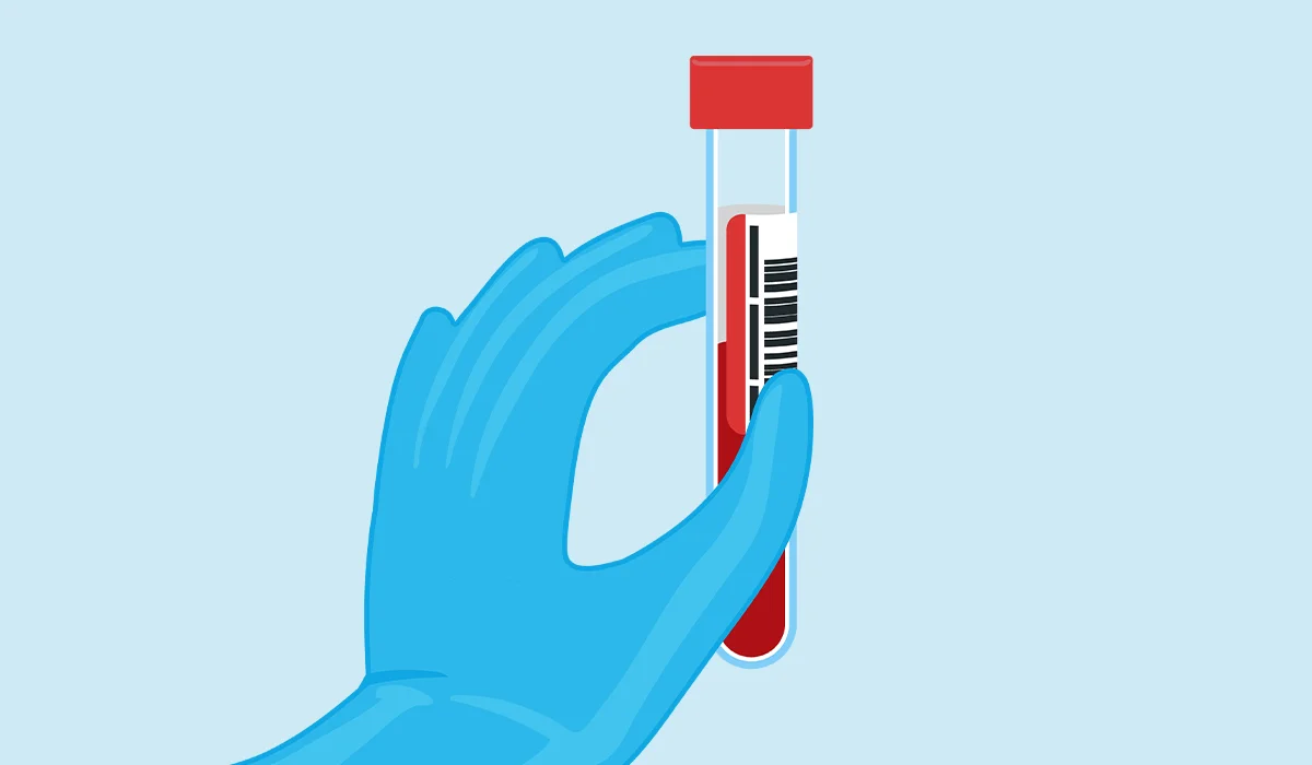
The basic diagnostic method for chromosome aberrations is karyotype testing. Karyotyping enables the detection of abnormalities in the structure and number of chromosomes. The most frequently used material for testing is venous blood from which the patient’s lymphocytes are isolated. Saliva, bone marrow, and fibroblasts can also be used as biological material. If it is necessary to perform a cytogenetic test on the fetus during pregnancy, amniotic fluid is collected.
There is no need to prepare for the test in advance, and the result is not influenced by the consumption of food, physical exercise, or the time spent collecting the material from the patient.
Karyotyping can be performed using three methods:
- Classic microscopic cytogenetic examination
- Fluorescence in situ hybridization (FISH)
- Comparative microarray hybridization techniques (aCGH)
Each cytogenetic test result should be consulted with a geneticist who will indicate further medical treatment.
Non-invasive prenatal tests (NIPT) provide an alternative to invasive procedures like the collection of amniotic fluid. These tests analyze free fetal DNA from a blood sample taken from the mother. Modern molecular biology approaches, including Next Generation Sequencing (NGS), such as the SANCO test, can be used to analyze the child’s genetic material. The NGS method is highly effective in detecting the presence of trisomy and monosomy in all 23 pairs of chromosomes, including chromosomes 21, 18, and 13. Importantly, this test is completely safe for both the mother and child.
When Should I Perform It?
Karyotype is not a routine test, and not all of us have performed it throughout our lives. However, there come times when they must be done.
A postnatal karyotype test, which involves examining the chromosomes in peripheral blood, is recommended in the following cases:
- When the patient displays physical features typical of a specific chromosomal syndrome, such as unusual eye spacing, a broad neck, or finger abnormalities (e.g., missing or extra fingers)
- If the patient has developmental issues, like a distinct heart defect, or experiences delays in psychomotor development
- In instances of repeated spontaneous miscarriages during the first trimester of pregnancy or if the patient has given birth to children with developmental problems
- When there are no signs of puberty, primary or secondary amenorrhea, and insufficient height in girls
- If there are abnormalities in the structure of the external genital organs
- In situations where a family member has a structural chromosome abnormality
Moreover, a bone marrow karyotype is conducted when suspected of a blood cancer disease, such as leukemia. This karyotype is related to cancer and is called an acquired karyotype.
A karyotype from amniotic fluid or trophoblast (the outer cell layer of the fetal membrane) is performed when fetal developmental defects are suspected, indicated by an abnormal ultrasound image and laboratory test results in a pregnant woman, and is called a prenatal karyotype.
Results
The results of the karyotype analysis may yield a normal outcome, typically denoted as 46, XX for females and 46, XY for males, or an abnormal outcome. An abnormal karyotype indicates structural abnormalities, which involve irregularities in the structure of the chromosomes (referred to as structural aberrations), or numerical abnormalities, where there is a deviation from the typical number of chromosomes (referred to as numerical aberrations).
Structural aberrations arise from the breakage and incorrect rejoining of one or more chromosomes. Structural aberrations include translocations, inversions, deletions, duplications, isochromosomes, and ring chromosomes. On the other hand, among numerical aberrations in humans, monosomies (the absence of one chromosome from a pair) and trisomies (an additional chromosome in a given pair) are the most prevalent.
Sources
- The Anatomy of a Chromosome. NIH.
https://www.ncbi.nlm.nih.gov/pmc/articles/PMC3284714/ - Genetics, Chromosomes. NIH.
https://www.ncbi.nlm.nih.gov/books/NBK557784/ - Chromosome Map. NIH.
https://www.ncbi.nlm.nih.gov/books/NBK22266/ - Exploring the Biological Contributions to Human Health: Does Sex Matter?. NIH.
https://www.ncbi.nlm.nih.gov/books/NBK222289/ - Genetics, Meiosis. NIH.
https://www.ncbi.nlm.nih.gov/books/NBK482462/ - The genetics of sex chromosomes: evolution and implications for hybrid incompatibility. NIH.
https://www.ncbi.nlm.nih.gov/pmc/articles/PMC3509754/ - Genetics, Chromosome Abnormalities. NIH.
https://www.ncbi.nlm.nih.gov/books/NBK557691/ - Genetic and Rare Disorders. NHS.
https://www.oxfordhealth.nhs.uk/cit/resources/genetic-rare-disorders/ - Down Syndrome. NIH.
https://www.ncbi.nlm.nih.gov/books/NBK526016/ - Edwards Syndrome. NIH.
https://www.ncbi.nlm.nih.gov/books/NBK570597/ - Patau Syndrome. NIH.
https://www.ncbi.nlm.nih.gov/books/NBK538347/ - Prader-Willi Syndrome. NIH.
https://www.ncbi.nlm.nih.gov/books/NBK553161/ - Angelman Syndrome. NIH.
https://www.ncbi.nlm.nih.gov/books/NBK560870/ - Williams Syndrome. NIH.
https://www.ncbi.nlm.nih.gov/books/NBK544278/ - Fragile X Syndrome. NIH.
https://www.ncbi.nlm.nih.gov/books/NBK459243/ - Genetic and genomic testing. NHS.
https://www.nhs.uk/conditions/genetic-and-genomic-testing/ - Genetics, Cytogenetic Testing And Conventional Karyotype. NIH.
https://www.ncbi.nlm.nih.gov/books/NBK563293/ - Non-Invasive Prenatal Testing (NIPT): Reliability, Challenges, and Future Directions. NIH.
https://www.ncbi.nlm.nih.gov/pmc/articles/PMC10417786/
